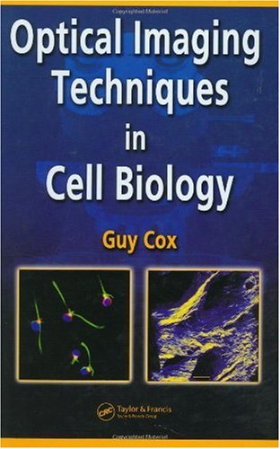
Since the word microscopy was coined in 1656, the evolution of the instrument has had a long and convoluted history. Plagued with problems of chromatic aberration, spherical aberration, and challenges with illumination and resolution, the microscope?s technical progression happened in a series of fits and starts until the late 19th century. After Ernst Abbe perfected the ?how? of lens design, achieving the theoretical limit imposed by wavelength, there came a revolution in subject matter or ?what? could be studied by microscope. Covering the entire field of microscopy, Optical Imaging Techniques in Cell Biology provides an overview of the technical evolution of the microscope and explains how the basics of optical microscopy led to the most advanced techniques employed today. The author addresses a vast array of topics including optical contrasting techniques, fluorescence, confocal versus widefield microscopes, lasers as a light source, and digital imaging, as well as the correction of aberrations that might arise. Building on this foundation, he then examines more advanced techniques such as quantitative fluorescence, fluorescence resonant energy, three-dimensional imaging, high-speed confocal microscopy, non-linear microscopy, and stimulated emission depletion. Delivering a truly comprehensive work encompassing the scope and breadth of the field, the author brings a new level of understanding to the student, technician, researcher, or investigator working in the fascinating realm of optical microscopy.


Reviews
There are no reviews yet.