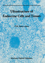
Innovative microscopic techniques, introduced during the last two decades, have contributed much to creating a new picture of the dynamic architecture of the cell, which can now be more exactly correlated with specific biochemical and physiopathological events. These developments have led to significant advances in our understanding of the physiomorphological and pathological aspects of the secretory mechanism, as well as the pharmacologic methods used to control, experimentally, the function of exocrine and endocrine glands. The integration of new ultrastructural methods such as freeze-fracture/etching, immunocytochemistry, scanning and high-voltage electron microscopy, cytoautoradiography, etc. , has proven to be of great value when applied to the study of endocrine cells and tissues. Because information on this topic has appeared in a variety of scientific and medical journals, this book: (1) reviews the results of an integrative approach presenting a comprehensive ultrastructural account of the main aspects of the field; (2) points out gaps or controversial topics in our knowledge; and (3) outlines pertinent directions for future research. The chapters, prepared by recognized authorities in the field, present traditional information on the topic in a concise manner and, with a valuable selection of original illustrations, show what the integration of new microscopic methods can contribute to the subject in terms of new concepts. This volume will be useful to cell biologists, anatomists, embryologists, histologists, pharmacologists, pathologists, and, of course, endocrinologists. It will also be of interest to students, practitioners of medicine, and to all others dealing with clinical research and diagnosis.
Biology
[PDF] Ultrastructure of Endocrine Cells and Tissues Kazumasa Kurosumi (auth.), P. M. Motta M.D., Ph.D. (eds.)
$19.99


Reviews
There are no reviews yet.